Surgical coordination of anterior cervical decompression interbody implant fusion
2024-05-23
Preoperative Preparation
Preparation of materials
1, disposable items: 20 #, 11 # blade, antibacterial surgical film, brain cotton pads, gelatin sponge, bone wax, electro-knife, bipolar electrocoagulation, suction tubes, suction head, 3-0, 4-0 (SL691) 7 * 17 triangular needles, 0, 1, 4, 7-gauge wires, absorbable threads, (if you take the skeleton bone, an additional antibacterial membrane)
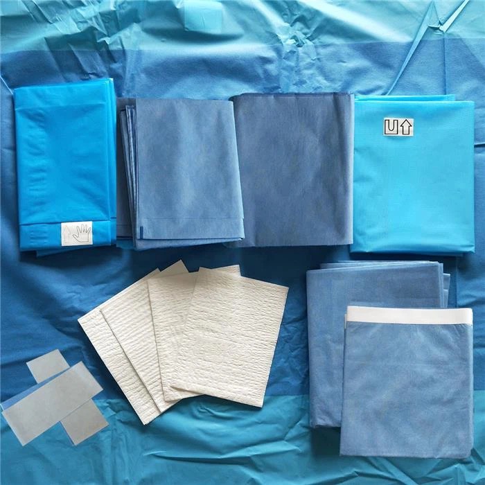
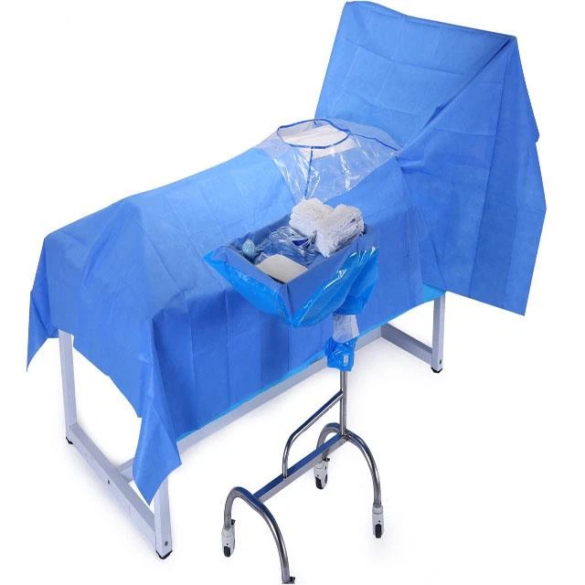
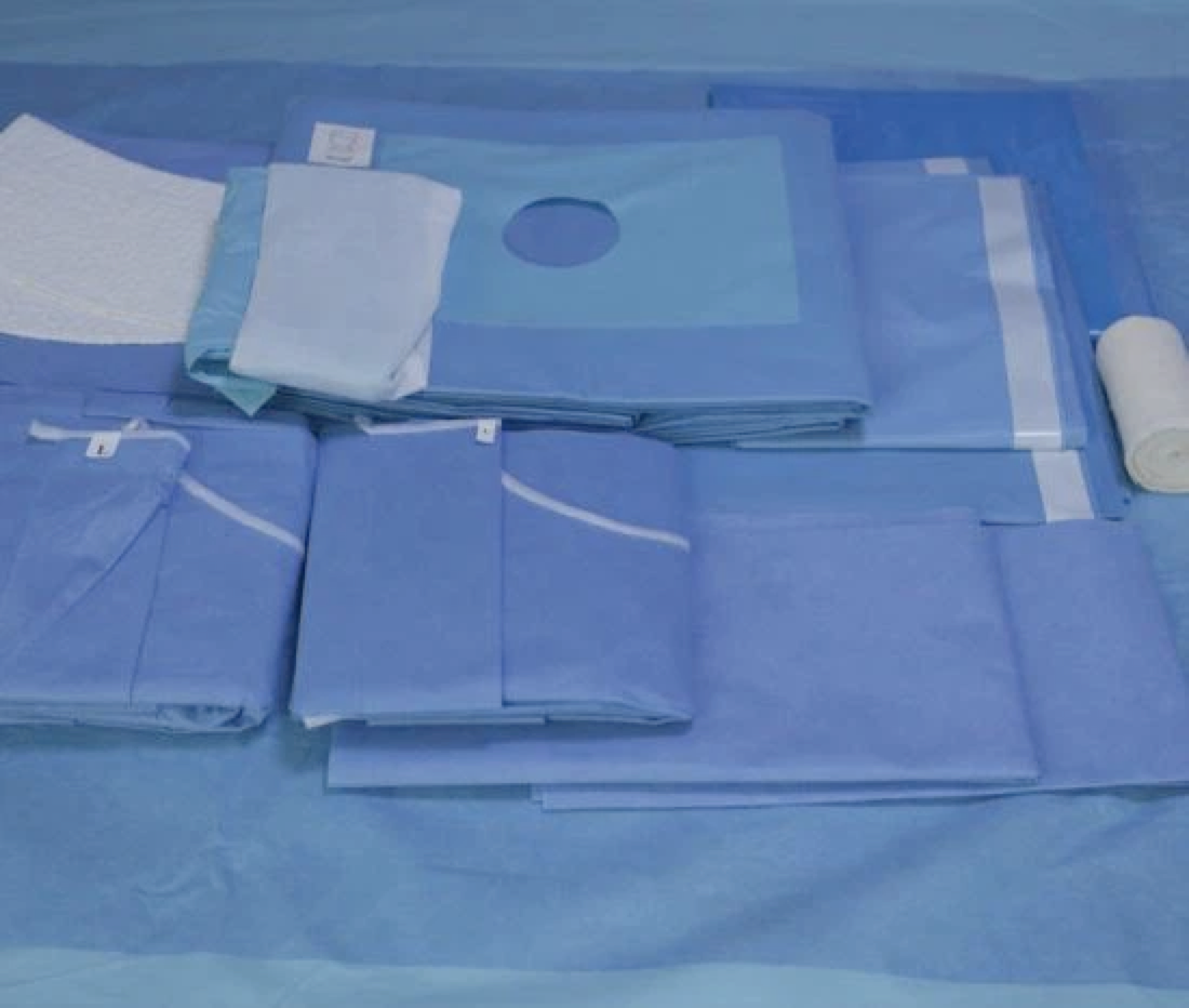
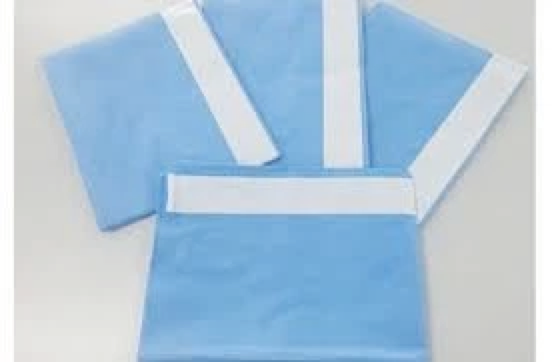
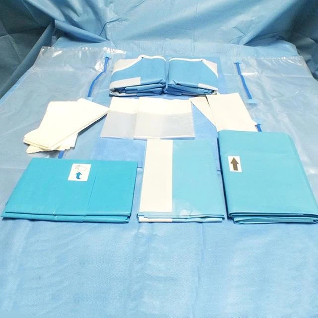
2, dressings: garment bag, single, dissection package, C-arm machine set
3, Instruments: vertebral plate package, anterior cervical instruments, manufacturers of instruments, the director of the special instruments
Anterior cervical spine surgery steps and surgical cooperation
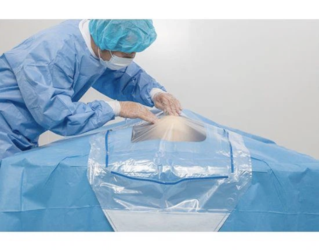
01 Sterilise and spread the towel, connect the electric knife and suction device. Passing knife, gauze, thyroid pulling hook, periosteal stripper;
02 Anterior cervical incision, separation of tissues, vascular forceps to separate the cervical latissimus dorsi muscle and reveal the anterior vertebral body
03 Placement of retractor stopper, propping up the surrounding soft tissues to fully expose the intervertebral space;
04 Pass the appropriate disc insertion and positioning needle, perform c-arm and x-ray machine fluoroscopy, pass gauze to fill in the wound and cover the surgical field with a sterile sheet;
05 Positioning drilling is performed on one vertebral body above and one below the diseased intervertebral space;
06 Installation of the anterior vertebral body support pegs;
07 Cut the anterior longitudinal ligament and pull it to both sides, pass 11# knife to cut, pass the cotton pads to compress the haemostasis, and suction to suck the blood (Note: the front end of the suction device can be set with a silicone urinary catheter of about 0.5cm in length, to prevent damage to the spinal cord);
08 Pass the 11# blade to remove the target intervertebral disc, pass the nucleus pulposus clamp to remove the excessive bone and soft tissue, fibrous annulus and nucleus pulposus, use the spatula, vertebral plate biting forceps and nucleus pulposus clamps to remove the excessive bone on the anterior wall and remove the bone cumbersome that grows anteriorly from the endplates (the removed bone is wrapped with gauze to preserve it for bone grafting);
09 Probe the posterior edge of the vertebral body for adhesions, and pass the nerve stripper to probe;
10 Start to install Cage trial mould: from small to large trial gap size, choose the appropriate Cage, fill the prepared bone block into the Cage, flush the vertebral body gap, between the vertebral body into the Cage;
11 Sometimes a columnar titanium mesh filled with bone is installed, gauze is used to fill the wound, a sterile medium sheet covers the surgical field, and a c-arm x-ray machine is performed for fluoroscopy;
12 Preparing the anterior cervical plate instrumentation and fixing the cage;
13 Set the appropriate length of plate and pre-curve it to conform to the curve of the cervical spine;
14 The plate is removed to the operator by hand-held forceps;
15 According to the anterior and posterior diameter of the cone, choose different lengths of screws and adjust the length of the insurance drill. (14 mm for men, 12 mm for women);
16 Insert the drill bit into the drilling sleeve and drill the hole according to the length of the screw;
17 Fill the wound again with gauze, cover the surgical field with a sterile mid sheet and perform a c-arm x-ray machine fluoroscopy;
18 Prepare to remove the nail, smear bone wax over the nail eye to stop bleeding, stop bleeding thoroughly, and place a drain. 3-0 absorbable suture of broad fascia to the subcutis, and 4-0 absorbable thread intradermal suture. The number and completeness of gauze, cotton instruments and sutures were counted and matched to the preoperative values;
Precautions
1. Position fixation. Patients with cervical spine injuries, regardless of preoperative and postoperative, should not move their necks when assisting with body positioning.
2. If autologous bone graft is taken, make sure to keep the removed bone and count the stitches and dressings at the bone removal site separately.
3. During the operation, keep the positioning needle, silicone urinary catheter head and cerebral tamponade, especially the small cut cerebral tamponade, the upper part of the table must be with silk thread, and check its integrity.
4. When installing the cage, pay attention to a timely reminder to remove the stuffed brain tamponade.
5. Remember the type and number of bone blocks and steel plates for internal fixation on the table and tell the visiting nurse in time.
 Email : sales@hnmedtech.com